Diagnostic imaging is an exciting, rapidly evolving sector of equine medicine. Technology is moving rapidly and we are able to visualize nearly every structure in a horse's body.
We can obtain highly detailed information that enables us to make specific diagnosis of problems, find these problems earlier, and treat the patient with more targeted therapies. Ultimately, this should allow us to keep horses leading healthy, pain-free lives for longer.
Radiology
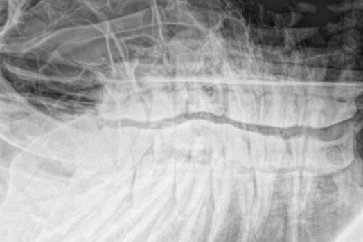
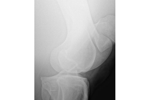
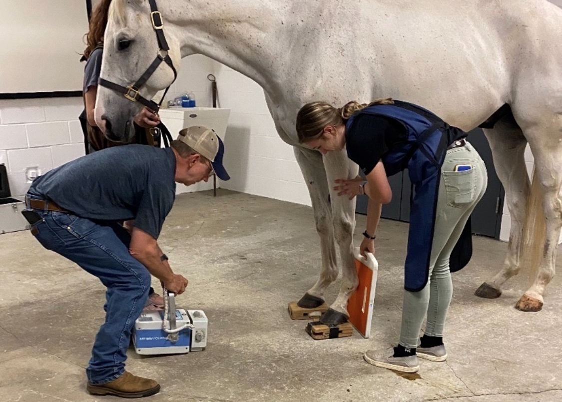
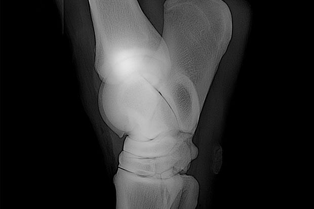
AVS Equine is proud to offer our clients direct digital x-rays. We have a 1000MgHz machine that allows us to view crisp x-rays of an adult horses entire skeleton. Our doctors can examine and optimize the image, along with providing the client a digital link. Our x-ray machines are powerful enough to penetrate an adult horse's chest, hips, and spine. This high-quality imaging allows doctors and clients to review results, diagnosis, and possibly treat in the course of one appointment.Digital radiography can be used to locate and evaluate a wide variety of irregularities in bone including abnormal bone appearance, early signs of degenerative change, and remodeling due to stresses.
Digital radiographs are invaluable as a screening procedure during lameness and pre-purchase exams to evaluate the general health of joints.
Ultrasound
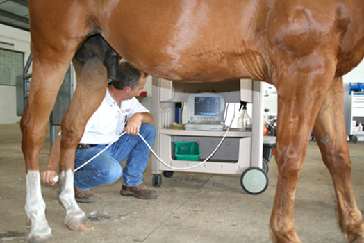
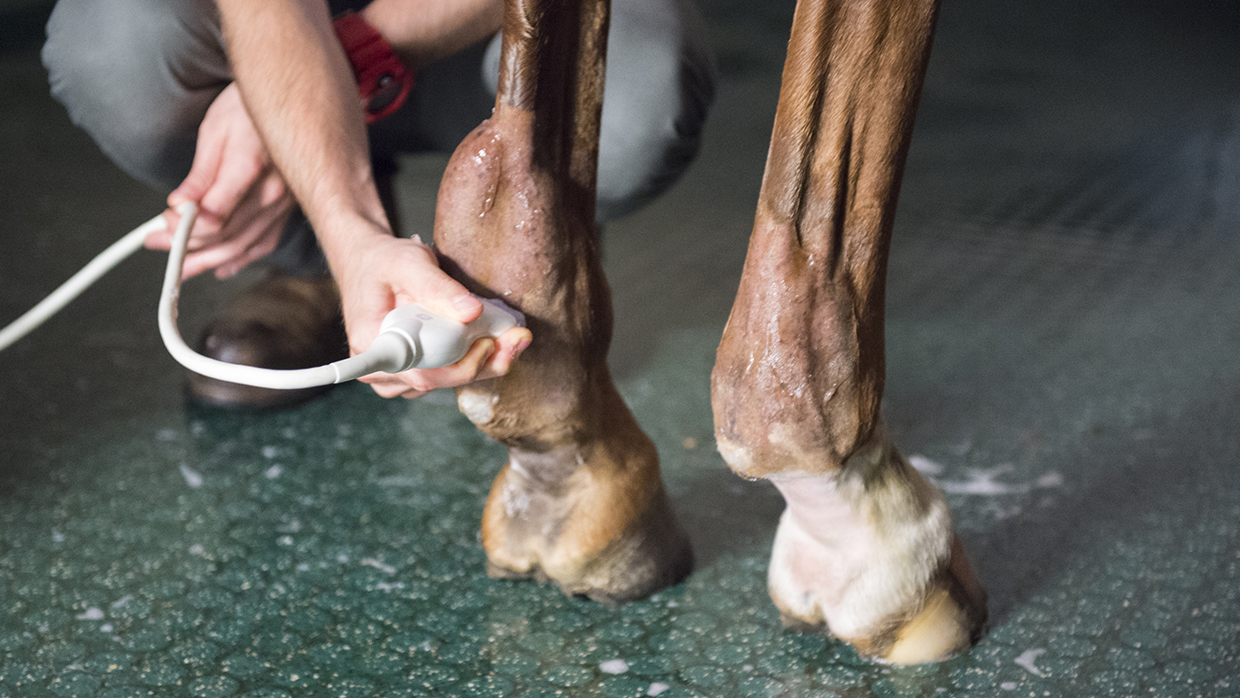
Sound waves are passed through the soft tissue and back to the transducer to allow for quick, harmless, and pain-free imaging of virtually any soft-tissue structure. Our 3MgHz to 10 MgHz digital ultrasound machines are used by our doctors to evaluate joint, tendon, and ligament injures and to monitor the healing progress. The digital ultrasound machine is also used by our doctors to evaluate pneumonia, abdominal pain, cardiac disease, umbilical infection, anatomical sites with a mass or swelling, late term pregnancy, and reproductive cycles.
Endoscopy
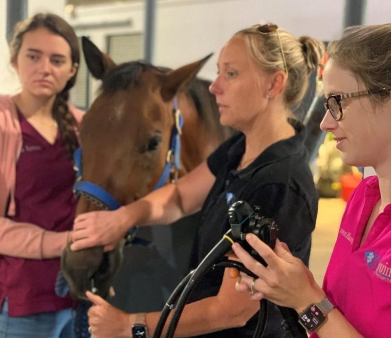
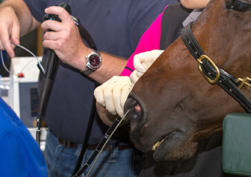
Endoscopy allows our doctors to see anatomical structures in the horse and is a key tool in the diagnosis of many conditions. Both a 1-meter and a 3-meter video endoscopy are available at AVS Equine Hospital and are commonly used to examine and evaluate the upper respiratory tract, upper gastrointestinal tract, urethra and bladder, as well as the uterus in the mare. The video endoscope is equipped with a monitor that allows the client to see these anatomical structures during the exam, which helps to strengthen the client's understanding of the nature of their horse's condition.
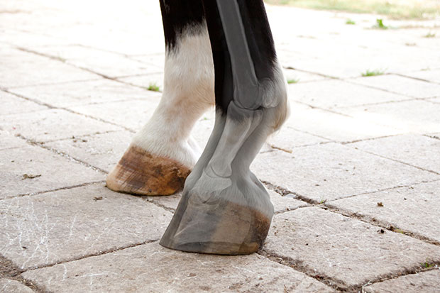
Computer Assisted Hoof Balancing
This state-of-the-art technology involves digital photos, digital radiographs and a computer program that measures and comp0ares several parameters to a database of thousands of sound horses. The doctor then works with the client and farrier to slowly bring the horse's hooves into optimum balance. Clients with competitive athletes have seen dramatic differences even after the first corrective measures.
